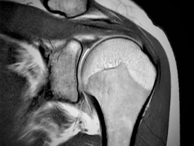
This module MRI of the shoulder – rotator cuff I provides steps for a structured approach for reporting the shoulder but with a focus on the rotator cuff. The rotator cuff is a challenging structure to assess with its complex and obliquely orientated anatomy.
In this module you will find different examples of rotator cuff tears and learn how to describe them, based on the recent and commonly agreed radiological literature.
While you go through the cases with the organized template you will become familiar and feel more confident with every next MRI of the shoulder. Several answers provide additional information on the how and why of the answers.
For your training several cases have been choses that may show limitations e.g. motion artefacts or suboptimal imaging planes as this is frequently the case in your clinical practice. Patients are in pain, or too large for regular positioning which brings on extra challenges to assess the relevant pathology. No perfect images from textbooks but daily practice defiances. We hope you’ll be able to deviate the challenge and learn going through the images
- Use a structured approach of an MRI of the shoulder
- Become familiar with the rotator cuff muscles and tendons
- Learn to detect and describe rotator cuff tears
- Become aware of possible associated pathology
Residents on year 4 and General radiologists, physiotherapists, orthopaedic surgery or general surgery
| Hardware | Tablets * | Minimum | Recommended |
|---|---|---|---|
| Memory (RAM): | 2 Gigabyte | 8 Gigabyte | 16 Gigabyte |
| Processor (CPU): | Dual core 1.85 Ghz | Dual core 2 Ghz | Quad core 2.5 Ghz |
| Internet connection | Minimum | Recommended | |
| Speed: | 10 Mbps | 25 Mbps | |
| Software | Tablets | Desktop | |
| Browser: | Safari * | Chrome ** | |
- * Tested with Safari on iPad 9.7 (2017), should also work on Android with Chrome. User interface not optimized for smaller screens. Large cases (more than 600 images) are not able to be opened on tablet or mobile devices due to memory consTableRowaints.
- ** Firefox, Edge and Safari also work but might not provide an equally smooth experience. Internet Explorer is not supported.


