This diagnostic imaging in lung cancer reporting course is aimed at radiologists, but also medical oncologists, radiation oncologists, and pulmonologists who are interested in improving their clinical skills about the interpretation and identification of images in the diagnosis, staging and response assessment of cases of lung cancer, principally using CT. Some cases with PET or MRI will also be shown.
The simulator enables practice in a real environment, working on cases of lung tumours. It is a “learning by doing” type of learning that allows you to learn from your errors and successes when interpreting the images. At the end of each case, the user will receive the answers immediately with comments both in the structured report and in the images, with the aim of reinforcing the entire learning process.
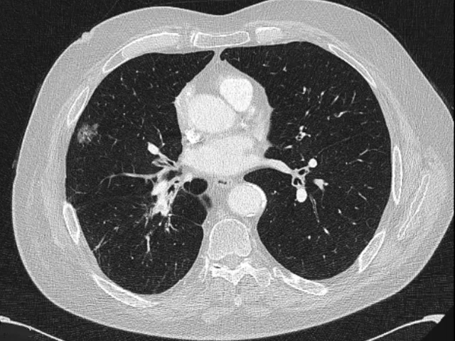
This thoracic radiology course contains 20 real anonymised cases selected by the mentor who prepared the content.
This medical training is organised by Unilabs Academy, an EACCME Trusted Provider.
Explore our Thoracic Radiology Fellowship Calendar 2026 — and take advantage of real‑time interaction with an expert mentor for personalised guidance, case discussions, and networking. Plus, stay up to date by learning the latest developments through focused, high‑impact lectures.
Testimonials:
"The simulator is a brilliant way to learn or relearn how to approach a certain examination. It allows you to practice, think logically through a case, and is very applicable to clinical work.”
Dr. Bryan Connolly, Specialist Radiologist
"I like everything and particularly the structured template which creates an algorithm of reporting”
Radiologist
How does it work?
The specialised radiologist mentor has carefully prepared a structured reporting template to help you report the cases, formatted in a fun clickable quiz format while you also view the cases in an web-based PACS-viewer on the right.
1. First, you should review the case images.
2. Then, you can begin the reporting template quiz (meanwhile still reviewing the images on the right)
3. After you submit your answers, the correct answers will appear and you will receive a score for your report.
4. Alongside the answers, you will also find helpful comments and learnings that the specialised radiologist mentor has left. There might also be arrows or markings on the images to help you see relevant findings.
5. If you score more than 66%, you have passed the module and can download your CME Certificate. If you did not pass, you can reset the module and try again.
You can save the module whenever you want and come back to it later, giving you flexibility to practice on your own time.
Don’t forget to watch introductory video to the right!
Don't miss our FAQ page for any questions you encounter along the way - or email us directly with your questions.
- Recognise the thoracic anatomy required to perform a correct TNM and staging classification for lung cancer
- Learn about the 8th edition of the TNM classification, its unique features and differences compared to previous versions
- Recognise the essential findings that can influence the various therapeutic options
- Learn the basics of response assessment using RECIST 1.1 and using immune response criteria
- Learn to identify patterns of pulmonary toxicity
| Hardware | Tablets * | Minimum | Recommended |
|---|---|---|---|
| Memory (RAM): | 2 Gigabyte | 8 Gigabyte | 16 Gigabyte |
| Processor (CPU): | Dual core 1.85 Ghz | Dual core 2 Ghz | Quad core 2.5 Ghz |
| Internet connection | Minimum | Recommended | |
| Speed: | 10 Mbps | 25 Mbps | |
| Software | Tablets | Desktop | |
| Browser: | Safari * | Chrome ** | |
- * Tested with Safari on iPad 9.7 (2017), should also work on Android with Chrome. User interface not optimized for smaller screens. Large cases (more than 600 images) are not able to be opened on tablet or mobile devices due to memory consTableRowaints.
- ** Firefox, Edge and Safari also work but might not provide an equally smooth experience. Internet Explorer is not supported.