This reporting CT course is aimed at the general radiologist and senior radiology registrar exposed to day-to-day high and low-impact trauma to the chest.
This exercise will equip the learner with the skills and knowledge necessary to interpret trauma CT chest scans using a systematic approach, ensuring that no critical injuries are missed.
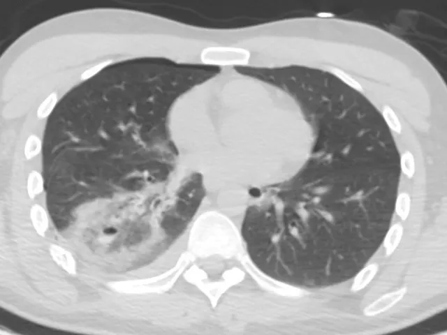
The course contains 10 real anonymised chest CT cases / 2 modules selected by the mentor.
By the end of each module, you will be able to:
1. Adopt a systematic approach at interpreting the trauma CT chest.
2. Describe common pathological entities acquired secondary to blunt and penetrating injuries to various parts of the thorax, namely:
- Pleura
- Lungs
- Mediastinum
- Chest wall
- Thoracic spine
CME Accreditation
This is a CME-accredited activity provided by Unilabs Academy, an EACCME Trusted Provider. Upon completion of each module, you will instantly receive 2 CME credits with a downloadable CME certificate.
Testimonials:
"The simulator is a brilliant way to learn or relearn how to approach a certain examination. It allows you to practice, think logically through a case, and is very applicable to clinical work.”
Dr. Bryan Connolly, Specialist Radiologist
How does Radiology Simulator work?
The specialised radiologist mentor has carefully prepared a structured reporting template to help you report the cases, formatted in a fun, clickable quiz format while you also view the cases in a web-based PACS-viewer on the right.
1. First, you should review the case images.
2. Then, you can begin the reporting template quiz (meanwhile still reviewing the images on the right)
3. After you submit your answers, the correct answers will appear and you will receive a score for your report.
4. Alongside the answers, you will also find helpful comments and learnings that the specialised radiologist mentor has left. There might also be arrows or markings on the images to help you see relevant findings.
5. If you score more than 66%, you have passed the module and can download your CME Certificate. If you did not pass, you can reset the module and try again.
You can save the module whenever you want and return to it later, providing you with flexibility to practice at your convenience.
- Adopt a systematic approach at interpreting the trauma CT chest.
- Describe common pathological entities acquired secondary to blunt and penetrating injuries to various parts of the thorax, namely: Pleura, lungs, mediastinum, chest wall, thoracic spine
| Hardware | Tablets * | Minimum | Recommended |
|---|---|---|---|
| Memory (RAM): | 2 Gigabyte | 8 Gigabyte | 16 Gigabyte |
| Processor (CPU): | Dual core 1.85 Ghz | Dual core 2 Ghz | Quad core 2.5 Ghz |
| Internet connection | Minimum | Recommended | |
| Speed: | 10 Mbps | 25 Mbps | |
| Software | Tablets | Desktop | |
| Browser: | Safari * | Chrome ** | |
- * Tested with Safari on iPad 9.7 (2017), should also work on Android with Chrome. User interface not optimized for smaller screens. Large cases (more than 600 images) are not able to be opened on tablet or mobile devices due to memory consTableRowaints.
- ** Firefox, Edge and Safari also work but might not provide an equally smooth experience. Internet Explorer is not supported.