This radiology reporting course includes 20 clinical stroke CT cases grouped in 4 training modules for you to report in our Radiology Simulator ( integrated online PACS viewer with structure reporting and immediate feedback with annotations and hangings).
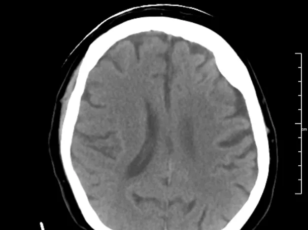
Stroke is one of the most common indications for brain imaging. Although the diagnosis of acute stroke is mainly clinical, imaging is often required to: rule out a contraindication for thrombolysis; differentiate cerebral infarction from haemorrhage; assess the extent of the stroke/complications, and rule out any other reasons for the acute presentation.
Although this course is based on on-call exposure to patients presenting with stroke, it will also show stroke at different stages.
Many of our cases are plain CT head scans; other techniques may be used when appropriate. We will also show variants that may mimic stroke and, when appropriate, provide a differential diagnosis for the findings seen on the images.
The course is delivered using a Reporting Simulator — a unique, interactive tool integrated in our e-learning platform designed for radiologists to mirror real-life clinical practice.
The Simulator includes a web-based DICOM image viewer and a dynamic, structured reporting form. Upon report submission, you receive immediate, case-specific feedback, allowing you to identify gaps and refine your reporting skills.
You can start for free and complete the first 5 cases ( plus 3 webinars!) at no cost.
Full access to all 20 cases in this course can be unlocked by purchasing a Premium Membership.
📅 Join also our CT-based Stroke Imaging online fellowship with Prof Johan Wasselius in December 2026.
Live lectures, case readings, and plenty of Q&A in an interactive format.
Check the agenda and schedule here
- To prepare the reporting radiologist to be able to give a confident diagnosis and to supply the appropriate information required for the appropriate management in cases of stroke.
- To keep the radiologist aware of the significant role an acute diagnosis can have on patient management.
- To enforce the importance of communication with the clinician, in particular, in case urgent intervention is required.
This course is geared towards senior residents and general radiologists wanting to increase their knowledge in neuroradiology, particularly in the area of stroke.
Radiology Simulator - How does it work?
The specialised radiologist mentors have carefully prepared a structured reporting template to help you report the cases, formatted in a quiz format while you also view the cases in a web-based PACS-viewer on the right.
1. First, you should review the case images.
2. Then, you can begin the reporting template quiz (meanwhile still reviewing the images on the right)
3. After you submit your answers, the correct answers will appear and you will receive a score for your report.
4. Alongside the answers, you will also find helpful comments and learnings that the specialised radiologist mentor has left. There might also be arrows or markings on the images to help you see relevant findings.
5. If you score more than 66%, you have passed the module and can download your CME Certificate. You can reset the module and try again if you did not pass.
You can save the module whenever you want and return to it later, giving you the flexibility to practice on your own time.
Check our FAQ page you can also email us directly with your questions.
| Hardware | Tablets * | Minimum | Recommended |
|---|---|---|---|
| Memory (RAM): | 2 Gigabyte | 8 Gigabyte | 16 Gigabyte |
| Processor (CPU): | Dual core 1.85 Ghz | Dual core 2 Ghz | Quad core 2.5 Ghz |
| Internet connection | Minimum | Recommended | |
| Speed: | 10 Mbps | 25 Mbps | |
| Software | Tablets | Desktop | |
| Browser: | Safari * | Chrome ** | |
- * Tested with Safari on iPad 9.7 (2017), should also work on Android with Chrome. User interface not optimized for smaller screens. Large cases (more than 600 images) are not able to be opened on tablet or mobile devices due to memory consTableRowaints.
- ** Firefox, Edge and Safari also work but might not provide an equally smooth experience. Internet Explorer is not supported.