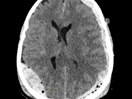
Traumatic Brain Injuries
Description
Traumatic brain injury is one of the most common causes of urgent imaging in busy emergency departments; every radiologist must have confidence in reading trauma head CT studies.
This radiology reporting course promotes a structured approach to interpreting traumatic brain studies, helping you produce a clear and consistent CT brain report while significantly increasing sensitivity to subtle injuries in otherwise near‑normal scans.
The course will demonstrate, through interactive examples, the common patterns of traumatic intracranial haemorrhages and the key points that help differentiate them, whenever possible. On the other hand, some cases were specifically selected to highlight less common trauma-related findings, which are important to be aware of and not overlook when reading studies in your practice.
The training contains real-life anonymised CT cases, drawn from everyday reporting in the on‑call department at TMC, Unilabs’ teleradiology company.
The course is delivered using a Radiology Simulator — a unique, interactive tool designed for radiologists to mirror real-life clinical practice. The simulator includes a DICOM image viewer and a dynamic, structured reporting form. Upon report submission, you receive immediate, case-specific feedback, annotated images allowing you to identify gaps and refine your reporting skills.
Accreditation and CME certificate
This is a CME-accredited radiology training by Unlibas Academy, an EACCME Trusted Provider. 2 CME credits will be granted upon completion.
Testimonials
💬 “The Unilabs radiology simulator is excellent. It’s intuitive, realistic, and a great tool for learning and practice. Dr Jonas Anuzis provides excellent, high-quality cases, along with valuable tips and constructive feedback. Excellent stuff!"
Dr. Jacques
📅 Looking for more advanced neuroradiology training?
Our live fellowships offer advanced, mentor-led training with extensive case review and structured guidance. Expect live lectures, hands-on reporting in a PACS environment, and interactive Q&A.
Explore also our accredited neuroradiology online fellowships with top European mentors - CME Calendar 2026 >
Learning Objectives
- Structural approach to brain trauma CT study reading
- Identifying and differentiating intracranial injuries and skull fractures
Level
The content will be relevant to anybody who works in the emergency radiology department.
Radiology Simulator - How does it work?
The specialised radiologist mentor - Dr Jonas Anuzis has carefully prepared a structured reporting template to help you report the cases, formatted in a quiz format while you also view the cases in a web-based PACS-viewer on the right.
1. First, you should review the case images.
2. Then, you can begin the reporting template quiz (meanwhile still reviewing the images on the right)
3. After you submit your answers, the correct answers will appear and you will receive a score for your report.
4. Alongside the answers, you will also find helpful comments and learnings that the specialised radiologist mentor has left. There might also be arrows or markings on the images to help you see relevant findings.
5. If you score more than 66%, you have passed the module and can download your CME Certificate. If you did not pass, you can reset the module and try again.
You can save the module whenever you want and return to it later, giving you the flexibility to practice on your own time.
If this is your first Radiology Simulator course, don't miss the introductory video to the right.
Technical Requirements
| Hardware | Tablets * | Minimum | Recommended |
|---|---|---|---|
| Memory (RAM): | 2 Gigabyte | 8 Gigabyte | 16 Gigabyte |
| Processor (CPU): | Dual core 1.85 Ghz | Dual core 2 Ghz | Quad core 2.5 Ghz |
| Internet connection | Minimum | Recommended | |
| Speed: | 10 Mbps | 25 Mbps | |
| Software | Tablets | Desktop | |
| Browser: | Safari * | Chrome ** | |
- * Tested with Safari on iPad 9.7 (2017), should also work on Android with Chrome. User interface not optimized for smaller screens. Large cases (more than 600 images) are not able to be opened on tablet or mobile devices due to memory consTableRowaints.
- ** Firefox, Edge and Safari also work but might not provide an equally smooth experience. Internet Explorer is not supported.
Introduction Video
Lecturers