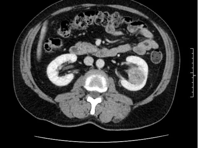
Kidney Cancer CT
Description
This kidney cancer imaging course is designed for general radiologists looking to strengthen their expertise in renal cell carcinoma (RCC) and renal mass interpretation using CT scan imaging. Through hands-on practice with real clinical cases, you will learn how to accurately identify kidney tumour patterns, perform imaging-based staging, and assess treatment response and follow-up. Working in a realistic reporting environment, you’ll build confidence in delivering clear, clinically relevant reports for kidney cancer management.
The training contains 10 anonymised real-life cases, grouped into 2 modules, drawn from the mentor's teaching files.
By achieving 66% correct answers in each module, the program will be accredited with 2 CME credits from the EACCME.
Unilabs Academy e-learning
The course is delivered using a Reporting Simulator — a unique, interactive tool integrated in our e-learning platform designed for radiologists to mirror real-life clinical practice. The Simulator includes a DICOM image viewer and a dynamic, structured reporting form. Upon report submission, you receive immediate, case-specific feedback, allowing you to identify gaps and refine your reporting skills.
You can start for free and complete the first 5 cases at no cost. Full access to all cases and other on-demand courses and webinars can be unlocked by purchasing a Premium Membership.
📽 If this is your first Radiology Simulator reporting course, don't miss the introductory video to the right!
Unilabs Academy short radiology fellowships
Continue your training with a live fellowship and join - Kidney imaging: From CT to MRI with Prof. Carlos Nicolau in Sept 2026. Live lectures, case reading, and plenty of Q&A in an interactive online format. Check the agenda and schedule here
Learning Objectives
- Recognise abdominal anatomy in order to perform a correct TNM classification and staging of the kidney tumours.
- Learn to identify the different types of renal tumours.
- Learn to classify kidney tumours using the indications set out in the 8th edition of the TNM classification for kidney tumours, paying close attention to the data in the image to be analysed.
- Recognise the essential findings that may influence the various therapeutic options.
Level
Technical Requirements
| Hardware | Tablets * | Minimum | Recommended |
|---|---|---|---|
| Memory (RAM): | 2 Gigabyte | 8 Gigabyte | 16 Gigabyte |
| Processor (CPU): | Dual core 1.85 Ghz | Dual core 2 Ghz | Quad core 2.5 Ghz |
| Internet connection | Minimum | Recommended | |
| Speed: | 10 Mbps | 25 Mbps | |
| Software | Tablets | Desktop | |
| Browser: | Safari * | Chrome ** | |
- * Tested with Safari on iPad 9.7 (2017), should also work on Android with Chrome. User interface not optimized for smaller screens. Large cases (more than 600 images) are not able to be opened on tablet or mobile devices due to memory consTableRowaints.
- ** Firefox, Edge and Safari also work but might not provide an equally smooth experience. Internet Explorer is not supported.
Introduction Video
Lecturers