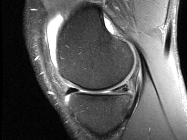
Meniscal Tear MRI
Description
This MSK online course helps general radiologists progress in their sub-specialisation. It focuses on common meniscal pathologies, injury mechanisms, and associated imaging findings, enabling learners to confidently describe meniscal tears and assess their stability in clinical practice.
The MRI training contains 25 anonymised real-life cases, grouped into 5 modules, drawn from the mentor's teaching files.
By achieving 66% correct answers in each module, the program will be accredited with 2 CME credits from the EACCME.
You can start for free and complete the first 5 cases at no cost. Full access to all cases and other on-demand courses and webinars can be unlocked by purchasing a Premium Membership.
Unilabs Academy e-learning
The course is delivered using a Reporting Simulator — a unique, interactive tool integrated in our e-learning platform designed for radiologists to mirror real-life clinical practice.
The Simulator includes a DICOM image viewer and a dynamic, structured reporting form. Upon report submission, you receive immediate, case-specific feedback, allowing you to identify gaps and refine your reporting skills.
Testimonials:
"The simulator is a brilliant way to learn or relearn how to approach a certain examination. It gives you the opportunity to practice, think logically through a case, and is very applicable to clinical work.”
Dr. Bryan Connolly, Specialist Radiologist,
"Very practical training tool which I have used frequently to practice. I love the step-by-step teaching method. Thank you!”
Dr. Anees Jwad, Radiologist,
Unilabs Academy mini fellowships.
Continue your MSK training with in-depth fellowships - live lectures, group case reading sessions, and plenty of Q&A with a mentor in an interactive online format.
Check the upcoming fellowship courses in 2026 here
Reporting Simulator - How does it work?
The specialised radiologist mentor has carefully prepared a structured reporting template to help you report the cases, formatted in a fun clickable quiz format while you also view the cases in a web-based PACS-viewer on the right.
1. First, you should review the case images.
2. Then, you can begin the reporting template quiz (meanwhile still reviewing the images on the right)
3. After you submit your answers, the correct answers will appear and you will receive a score for your report.
4. Alongside the answers, you will also find helpful comments and learnings that the specialised radiologist mentor has left. There might also be arrows or markings on the images to help you see relevant findings.
5. If you score more than 66%, you have passed the module and can download your CME Certificate. If you did not pass, you can reset the module and try again.
You can save the module whenever you want and come back to it later, giving you flexibility to practice on your own time.
If this is your first Radiology Simulator course, don't miss the introductory video to the right!
Check our FAQ page you can also email us directly with your questions.
Learning Objectives
- To know and correctly describe a meniscal tear
- To be able to recognize indirect signs
- To learn how to assess the stability of a meniscal tear
- To become familiar with a common injury mechanism involving the menisci
Level
Technical Requirements
| Hardware | Tablets * | Minimum | Recommended |
|---|---|---|---|
| Memory (RAM): | 2 Gigabyte | 8 Gigabyte | 16 Gigabyte |
| Processor (CPU): | Dual core 1.85 Ghz | Dual core 2 Ghz | Quad core 2.5 Ghz |
| Internet connection | Minimum | Recommended | |
| Speed: | 10 Mbps | 25 Mbps | |
| Software | Tablets | Desktop | |
| Browser: | Safari * | Chrome ** | |
- * Tested with Safari on iPad 9.7 (2017), should also work on Android with Chrome. User interface not optimized for smaller screens. Large cases (more than 600 images) are not able to be opened on tablet or mobile devices due to memory consTableRowaints.
- ** Firefox, Edge and Safari also work but might not provide an equally smooth experience. Internet Explorer is not supported.
Introduction Video
Lecturers