
On-demand Webinar
·
1 CME Credit
Abdominal injuries: the role of computed tomography
Trauma CT protocols support detection of blunt, penetrating, and blast-related abdominal injuries.
Already have an account?
Description
Contrast-enhanced CT strategy is examined in relation to diagnostic delay in abdominal trauma. The focus includes bowel, diaphragmatic, pancreatic, abdominal-wall, penetrating, and blast injuries, radiological signs of hypovolaemia, and challenges that cause injuries to be missed.
Learning Objectives
- Apply an appropriate contrast-enhanced CT protocol for abdominal trauma.
- Recognise bowel, diaphragmatic, pancreatic, and abdominal-wall injuries.
- Assess penetrating and blast trauma and identify CT signs of hypovolaemia.
Topics Covered
To jump to a specific chapter, click on the chapter title once the video is playing.
00:00 - Introduction and Importance of Abdominal Trauma Imaging
01:13 - Clinical Challenges in Diagnosing Abdominal Injuries
02:12 - Imaging Techniques for Abdominal Trauma
03:14 - CT Scanning Protocols for Trauma
07:54 - The Use of IV Contrast in Imaging
19:37 - Challenges with Diagnosing Bowel Injuries
21:58 - Diaphragmatic Injuries and Their Complexities
24:54 - Pancreatic and Abdominal Wall Injuries
28:51 - The Complexity of Penetrating Wounds
32:52 - Imaging and Managing Blast Injuries
33:56 - Radiological Signs of Hypovolemia
35:54 - The Role of Imaging in Reducing Trauma Mortality
46:34 - Q&A Session with Experts
Lecturers
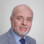
András PalkóM.D. Ph.D.
Szeged, Hungary
Consultant radiologist at TMC and professor of Radiology at University of Szeged, Hungary
Professor András Palkó, M.D, Ph. D. has extensive clinical as well as academic experience. Dr. Palkó has been working with abdominal CT and MRI for 25 years. He is the author of 90 peer reviewed papers and over 40 books and book chapters, invited speaker of international scientific and educational conferences, co-founder and faculty member of the European School of Radiology and examiner for the European Diploma in Radiology



